Products
HSA SCAN
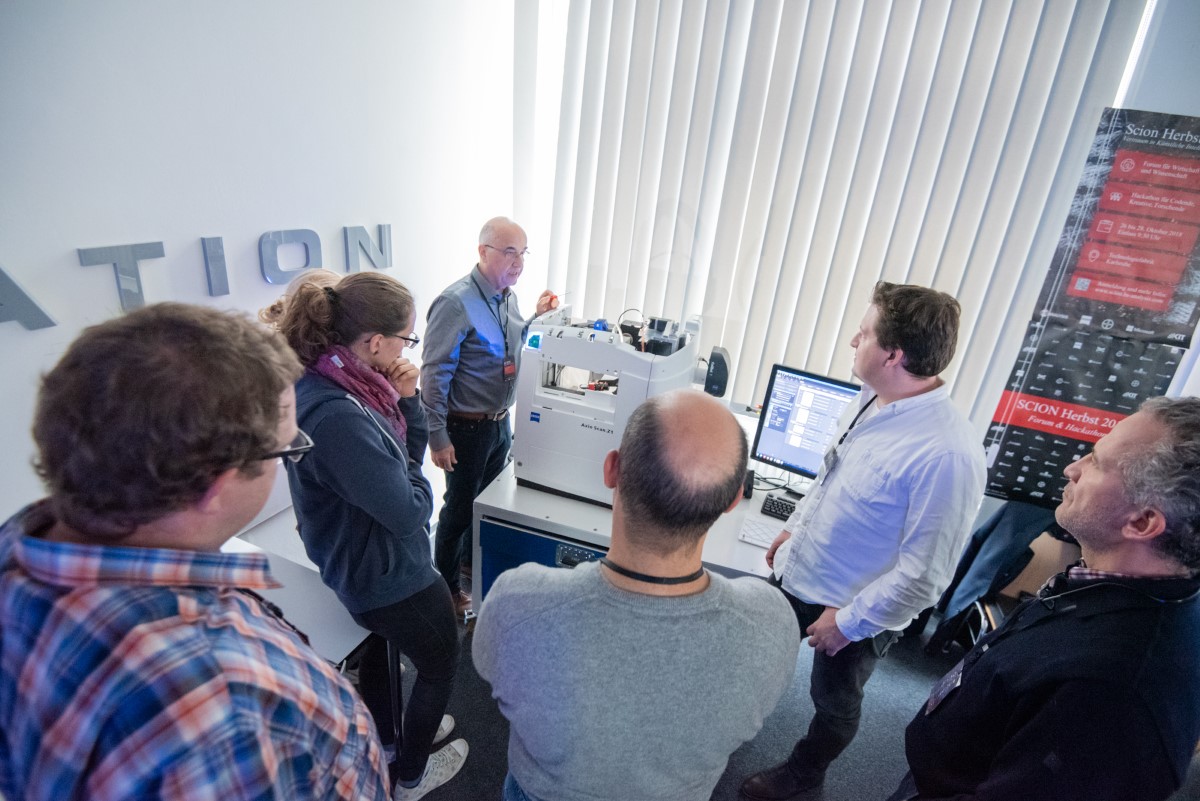
With HSA SCAN, analog data is transformed into digital formats. HS Analysis is one of the world’s leading companies, focusing on precise and efficient digitization technologies. The areas of application are diverse, ranging from industry and document management to the biomedical field. Cameras are mounted on racks or microscopes, enabling manual, semi-automated, or fully automated data digitization.
HSA Consulting
HS Analysis is a global leader in consulting for digitization and the integration of AI. We specialize in supporting our customers in solving their unique image analysis challenges—whether through traditional methods or advanced AI-driven solutions. Our expert team is dedicated to thoroughly analyzing your specific requirements and helping you choose the right tools and workflows. Through personalized consulting, we ensure that you receive the perfect solution to maximize efficiency and achieve your desired results. Let HSA Consulting be your partner in unlocking the full potential of your image analysis projects.
HSA STUDY
HSA STUDY is management software for the sustainable and reproducible execution of studies. The focus is on the personnel-independent collection of large data sets, the corresponding deep learning models, and ultimately the results.
HSA KIT
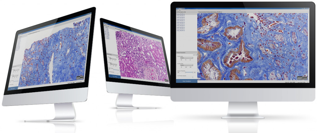
With HSA KIT, managing large-scale projects has never been easier. You can create projects containing as many files as you need and initiate analysis for all of them with a single click. HSA KIT automatically handles the entire process, analyzing each file independently, allowing you to focus on results rather than manual execution. Simplify your workflow and save time with HSA KIT’s efficient batch analysis capabilities.
HSA GTD Service
Purchase the HSA GTD Service. Experts at various levels create annotations according to your specifications, supported by deep learning professionals to ensure high-quality annotations. GTDs can be created for images, sound, video, text, invoices, Excel data, and measurement data. Annotations are done using the HSA KIT Module Annotator, which allows the export of GTDs in any format and ensures seamless integration into other software solutions.
HSA Annotator
HS Analysis is the global leader focused on providing professional annotation tools for various data formats. The HSA Annotator is a module in HSA KIT and is a powerful tool for quickly and professionally creating high-quality training data. It enables the annotation of images, sound, videos, texts, invoices, Excel data, and other measured signals. Data can be archived and tracked for future training iterations, ensuring independence from the annotators. A protocol for standardized annotations is also provided by HSA. All annotations can be exported in common formats and integrated into other software solutions. APIs are also available.
HSA FILE Viewer
The HSA FILE Viewer is the ideal solution for searching and opening data directly from the hard drive or a database. Various data types, such as spectra, acoustic signals, or image sequences, can be filtered and displayed precisely according to your needs. The HSA FILE Viewer is designed to professionally handle specific formats, such as spectral data or WSIs, offering optimal channel separation and efficient pyramid display. HS Analysis is one of the global leaders, focusing on advanced solutions for professional viewing tools.
HSA CASES
HS Analysis is the market leader in the DACH region, focusing on innovative software solutions for managing clinical cases. HSA CASES helps doctors structure, filter, and maintain an overview of patient data in their daily clinical routine. Thanks to seamless integration with medical infrastructure, cases can be imported, edited, and saved from existing systems. HSA CASES also provides an optimal interface for the future integration of AI into everyday clinical practice.
HSA DASH
Elevate your hospital’s operational efficiency and patient care with HS Analysis’ advanced dashboard solutions. Unlike traditional Hospital Information Systems (HIS), our intuitive and user-friendly dashboards seamlessly integrate with your existing systems, providing real-time, actionable insights that drive informed decision-making across departments. Our solutions are designed to enhance collaboration, streamline data access, and scale with your hospital’s evolving needs.
HSA DASH enables doctors to filter patient groups and visualize the progress of treatments and medications. It also tracks the timeline of a specific patient’s treatment, which can be shared and discussed with colleagues through interactive graphs. Over time, this helps doctors monitor improvements or deteriorations under certain therapies. HSA DASH is also a future-proof interface for AI integration, enabling the prediction of treatment outcomes for new patients.
HSA API
HSA Analysis products offer open interfaces that allow any function to be integrated into external systems through APIs. This ensures seamless integration of digitization services, the export or import of training data or AI models, and connectivity with existing infrastructures. Whether it’s LIS, KIS, dedicated servers, or cloud solutions like AWS, HSA API is flexible and adaptable to your needs.
HSA AI Cockpit
HSA PRED FC
HSA PRED FC stands for Prediction of Fabrication Conditions. To ensure sustainable and reproducible results in laboratories, labs use HSA PRED FC to enter all input parameters of fabrication processes for materials into the software and over time check which changes in the composition of the fabrication parameters are necessary to achieve the desired material quality. In parallel, AI evaluates the composition of the parameters and makes its own suggestions.
HSA Academy
HSA Academy offers tailored training programs that help medical professionals expand their skills in using modern technologies such as programming and artificial intelligence. Our hands-on courses prepare you optimally for the application of these innovative technologies in healthcare. Upon completion, participants receive a certification that validates their newly acquired competencies.
HSA Courses
HS Analysis provides specialized training for medical professionals to learn programming languages like R and Python, with a focus on deep learning applications. Doctors can learn to collaborate with ChatGPT for an interactive, efficient learning experience. Upon completion, participants will receive certification in programming skills.
HSA Workshops
Discover the world of Artificial Intelligence with HSA Workshops by HS Analysis! Our workshops combine engaging lectures from leading experts with interactive, hands-on exercises. You’ll have the opportunity to work with HSA specialists on real-world challenges and develop innovative solutions, particularly with the support of modern AI technologies. Benefit from our expertise and learn how to effectively leverage AI for your specific needs.
For pre- and clinical research and diagnostics, it is important that medical images are not just images, diagnostic reports are not just unstructured information, but also that molecular data is automatically evaluated and not separated from the results of image and text analysis.
This has been the expertise of HS Analysis GmbH since 2015. The autonomous AI systems support researchers and doctors in the automated quantification and prediction of medical data. This allows researchers and doctors to use their expert knowledge more efficiently and focus on research or disease. When it comes to automated image analysis, knowledge from publications and reports and the analysis and prediction of molecular data, the integration of the corresponding AI into the hardware and infrastructure is one of the main competencies of HS Analysis GmbH.
The company works closely with various partners worldwide when digitalization and automation are part of the daily routine in the lab or hospital. With the help of its own deep learning architecture, active and transfer learning, HS Analysis sets the global standard in diagnostic prediction, for example CDx and the early detection of rare diseases or types of cancer.
Modules
Ophthalmology
The field of ophthalmology is dedicated to the comprehensive study and management of eye diseases and disorders. It encompasses a range of diagnostic techniques and treatments aimed at preserving and improving visual health. Accurate and timely diagnosis is crucial, as early intervention can significantly impact patient outcomes. HS Analysis GmbH’s innovative modules leverage advanced AI algorithms to analyze ocular images with high precision. These AI-driven tools enhance the detection of various eye conditions, such as retinal diseases, glaucoma, and macular degeneration, by improving image clarity, automating anomaly detection, and providing detailed insights that assist clinicians in making informed decisions.
Ophthal Tumor Classifier
The Module Ophthal Tumor Classifier uses pre-trained models to accurately detect and classify ocular tumors in medical images.
By leveraging advanced deep learning algorithms, this module distinguishes between tumor and non-tumor regions based on characteristics such as size, shape, and location.
It enables early detection and precise analysis, aiding ophthalmologists in making informed decisions for effective treatment.
The module’s high accuracy and precision in identifying tumor activity contribute to improved patient outcomes and a reduction in morbidity and mortality.
Neuro Keratopath
The Module Neuro Keratopathy uses pre-trained models to accurately segment and classify corneal nerve structures in patients with neurotrophic keratopathy (NK).
By leveraging deep learning, the module processes IVCM images to detect significant reductions in nerve density, changes in epithelial and endothelial cell densities, and increases in dendritic immune cells.
This advanced tool aids ophthalmologists in diagnosing NK early, assessing disease severity, and monitoring treatment response, providing crucial insights for better patient outcomes.
Neuropathology
Neuropathology focuses on understanding and diagnosing diseases that affect the nervous system, including the brain, spinal cord, and peripheral nerves. This field involves the examination of neural tissues to identify pathological changes associated with neurological disorders such as Alzheimer’s disease, Parkinson’s disease, and multiple sclerosis. Accurate analysis of tissue samples is essential for effective diagnosis and treatment planning. HS Analysis GmbH offers sophisticated modules that utilize AI to enhance the analysis of neural tissue images. These AI-powered tools help in identifying subtle pathological features, improving diagnostic accuracy, and supporting cutting-edge research in neuropathology by providing deeper insights into the mechanisms of neurological diseases.
Neuro-COVID-19 and Alzheimer’s Disease
Through the proteolytic processing of amyloid precursor protein and the subsequent hyperphosphorylation of tau protein, Alzheimer’s disease leads to the accumulation of beta-amyloid peptides in the medial temporal lobe and neocortical structures, the most affected areas of the brain. This is one of the main causes of a series of events that result in mitochondrial dysfunction, synaptic dysfunction, and oxidative stress. Acute COVID-19 infections, which lead to death, occur more frequently in patients with Alzheimer’s disease.
Creutzfeldt-Jakob Disease (CJD) and Alzheimer’s Disease
In both Creutzfeldt-Jakob disease and Alzheimer’s disease, severe synaptic dysfunction occurs due to the misfolding of prion proteins in CJD and the accumulation of beta-amyloid peptides in Alzheimer’s. This leads to a progressive loss of nerve cells and, ultimately, irreversible dementia. In CJD, particular attention is given to the mechanisms by which pathological prions exert their toxic effects and accumulate in the brain.
Nephropathology
Nephropathology is concerned with the study of kidney diseases and disorders, focusing on the pathology of renal tissues. Effective diagnosis and management of kidney-related conditions, such as glomerulonephritis, diabetic nephropathy, and kidney tumors, require detailed examination of tissue samples. HS Analysis GmbH provides specialized modules that harness AI to analyze kidney tissue images with precision. These modules aid in the detection and classification of renal abnormalities by enhancing image quality, automating diagnostic processes, and offering detailed analytical reports that support both clinical diagnostics and research in nephropathology.
Kidney Analysis
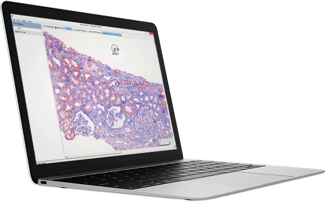
The “Kidney Analysis” Module is a powerful tool designed for precise kidney tissue analysis. Leveraging advanced pre-trained AI models, this module automatically identifies and segments key structures within whole slide kidney images, including Glomeruli and Tubuli. Ideal for research and diagnostics, it streamlines workflow, enhances accuracy, and provides valuable insights into kidney pathology with unparalleled efficiency.
Drug Development
Drug development is a complex and multi-stage process that involves the discovery, testing, and refinement of new medications. This process requires rigorous analysis and evaluation of various data types to ensure the efficacy and safety of drug candidates. While traditional methods often rely on imaging and biochemical assays, HS Analysis GmbH offers innovative modules that use Electronic Spectroscopy and Resonance (ESR) to analyze sound data. These modules provide valuable insights into the properties and behaviors of drug compounds, supporting the optimization of drug formulations and the evaluation of their performance throughout the development stages.
Vessel Detector
The Vessel Detector Module is a powerful tool that utilizes pretrained models to accurately segment blood vessels in images.
Beyond mere detection, this module further segments the vessels into tunica and lumen, providing detailed anatomical insights.
This precise segmentation is invaluable for a range of applications, including the study of vascular diseases, assessment of blood vessel integrity, and analysis of conditions such as atherosclerosis.
By offering detailed structural information, the Vessel Detector Module supports advanced research and clinical diagnostics, aiding in the development of targeted treatments and interventions.
gH2Ax Analyzer
The gH2Ax Analyzer module employs pre-trained AI models for segmentation, focusing on gH2Ax signals.
It processes these signals to generate comprehensive result tables, providing critical data for analyzing cellular responses to DNA damage.
This module supports precise therapy decisions and aids in the development of personalized treatment plans.
Lung Cancer Analysis
The Lung Cancer Analysis module utilizes pre-trained AI models for segmentation of Proximity Ligation Assay (PLA) signals in Non-Small Cell Lung Cancer (NSCLC). It processes these signals to generate detailed result tables, offering crucial insights into drug resistance. This enables more informed therapy decisions, particularly in tailoring personalized treatment strategies for lung cancer patients.
Membran Detection
The Membran Detection Module is a specialized tool for analyzing cell membranes in brightfield images using advanced pretrained models.
This module excels at detecting and identifying tumor and necrotic areas alongside the cell membranes. Additionally, it classifies cells based on their staining intensity, providing detailed insights into cellular characteristics.
The analysis culminates in the calculation of the H-Score, a crucial metric for evaluating staining patterns and intensities.
This module is indispensable for researchers and clinicians focused on cellular pathology and tumor analysis.
Dermatology
Dermatology focuses on the diagnosis and treatment of skin conditions and diseases, ranging from common issues like acne and eczema to more severe conditions such as skin cancer. Accurate diagnosis often involves the detailed examination of skin images to identify and assess various dermatological abnormalities. HS Analysis GmbH’s modules in dermatology utilize advanced AI to analyze skin images with high precision. These AI-driven tools assist in detecting and classifying skin conditions, enhancing the accuracy of diagnoses, and providing ongoing monitoring capabilities that support effective treatment planning and research into skin health.
CD11b Analyzer
The CD11b Analyzer Module is a specialized tool designed for detecting and analyzing cells stained with CD11b and DAPI in fluorescence images, particularly within large Whole Slide Images (WSIs).
This module performs precise image analysis to identify cells marked by the CD11b and DAPI stains, generating a comprehensive result table that includes the number and area of the detected cells.
This quantitative data is crucial for studies focusing on immune cell populations, such as macrophages and microglia, and their roles in various diseases, making the CD11b Analyzer an essential resource for immunological research and diagnostics.
ERG Analyzer
The module ERG Analyzer uses pre-trained models to segment and analyze ERG (endothelial cells) in multichannel fluorescence images, particularly within the context of skin cancer research.
By accurately identifying and quantifying ERG markers, this module provides critical insights into angiogenesis and tumor microenvironment interactions.
Integrated with the HSA KIT software, it facilitates standardized analysis, enabling quick and efficient processing of multiple medical images, ultimately aiding in the early detection, diagnosis, and treatment planning of conditions like melanoma.
This tool enhances the precision and productivity of research and clinical workflows.
Psoriasis Detection
The Psoriasis Detection Module is an advanced tool designed to analyze standard smartphone images for the presence of skin and psoriasis plaques using pretrained models.
This module not only detects plaques but also classifies them based on thickness, redness, and scaling using additional models.
The comprehensive analysis provided by this module enables the accurate calculation of the Psoriasis Area and Severity Index (PASI) Score, offering a valuable resource for both clinicians and patients to monitor and assess the severity of psoriasis.
Molecular Analysis
Our Molecular Analysis solutions provide advanced tools to investigate biological systems at the molecular level. By offering high-throughput and data-rich methodologies, we enable researchers and clinicians to gain deep insights into complex processes. Whether for research or clinical applications, our platform delivers the precision and flexibility needed to push the boundaries of discovery and improve medical outcomes.
HSA API
HS Analysis GmbH’s HSA KIT API is your ultimate solution for creating and managing ground truth data seamlessly. The API offers robust features including secure user authentication using JSON web tokens (JWT), comprehensive project management, efficient file handling, and real-time annotation updates. It integrates effortlessly into existing systems like Laboratory Information Systems (LIS) and Hospital Information Systems (HIS).
Colorectal Analysis
The Module Colorectal Analysis uses pre-trained models to accurately detect and classify colorectal cancer cells within medical images.
Leveraging advanced AI algorithms, this module identifies and categorizes different types of glands, distinguishing healthy tissues from adenoma and carcinoma glands.
Integrated within the HSA KIT software, it enables comprehensive data extraction, detailed reporting, and precise analysis, significantly aiding in the early detection and treatment of colorectal cancer, ultimately improving patient outcomes.
Annotator
The HSA Annotator is a module in HSA KIT and is a powerful tool for quickly and professionally creating high-quality training data. It enables the annotation of images, sound, videos, texts, invoices, Excel data, and other measured signals. Data can be archived and tracked for future training iterations, ensuring independence from the annotators. A protocol for standardized annotations is also provided by HSA. All annotations can be exported in common formats and integrated into other software solutions. APIs are also available.
Liver Analyzer
The module Liver Analyzer uses pre-trained models to segment liver tissue into two key structures: vessels and non-vessels.
By accurately identifying blood vessels and surrounding tissues within liver images, this module provides detailed insights into the liver’s vascular network and overall tissue composition.
This advanced segmentation tool is essential for analyzing liver health, diagnosing potential abnormalities, and supporting research efforts in hepatology.
Integrated within the HSA KIT software, it ensures precise, reproducible results, enhancing the quality of liver studies.
Heart Analyzer
The module Heart Analyzer uses pre-trained models to segment rat heart sections, specifically targeting cardiomyocytes in brightfield images.
By leveraging advanced deep learning techniques, this module accurately identifies and delineates cardiomyocyte structures, facilitating detailed analysis and enabling researchers to better understand heart tissue morphology and related conditions.
This tool is essential for conducting precise and efficient cardiac studies, supporting advancements in cardiovascular research and treatment.
CD3 – CD8 Analyzer
The CD3 – CD8 Analyzer Module is a specialized tool designed for the detection and analysis of CD3 and CD8 stained cells in fluorescence images, particularly within large Whole Slide Images (WSIs).
By performing image analysis on these massive datasets, the module accurately identifies cells stained with the corresponding markers and generates a detailed result table.
This table includes the count and area of the detected cells, providing essential quantitative data for immunological research and diagnostics.
This module is particularly valuable in studies of immune response and tumor microenvironments, where precise cell identification and quantification are critical.
ESR Training
The ESR Training module in HSA KIT allows researchers to create principal component analysis (PCA) models from known ESR spectra.
These models form a database for identifying and quantifying unknown substances in future samples.
By analyzing spectral patterns and correlating them with specific chemical properties, this module supports precise and reliable analysis, essential for material research, quality control, and compliance with industry standards.
ESR Evaluation
The ESR Evaluation module in HSA KIT is designed to assess unknown ESR spectra by comparing them with PCA models generated in the ESR Training module.
This process enables accurate identification and quantification of substances, with pass/fail indicators based on the degree of spectral match.
The module streamlines analysis, ensuring consistent, reliable results that support rigorous quality control and regulatory compliance across various industries.
Technology
Technology
Autonomous AI systems as stand-alone software or software in the lab’s LAN or in the cloud
Areas of application
Image analysis, molecular analysis and natural language processing as Data Science
Projects & Services
Multi-channel fluorescence, brightfield and EM data combined with NGS, PCR and NMR data and DB and text data
Examples
Cell tracking and analysis in real time
HSA internally developed deep learning
Tracking changes in cellular morphology and behavior in coordination with the surrounding cells. Cells with increasing area are marked in blue, cells with decreasing area in red. Length of individual cell borders, perimeter, area and percentile change compared to the previous image (Δ area, Δ perimeter) are tracked in real time.
Standard Deep Learning U-Net
Simple intensity-dependent annotation of the cell boundaries compared to the original image.
AI networks developed in-house:
- + Cell border gaps in the original image are closed intelligently
- + Automatically track the length of individual cell boundaries, changes in cell morphology and behavior in coordination with surrounding cells
- + Visual representation of selected morphological changes (area, circularity, behavior of neighboring cells, etc.)
- + Simple export of all statistical data to an Excel file
Old U-Net based AI network:
- – Gaps in the original image are not closed, which affects the subsequent statistical analysis
- – Tracking of cellular parameters difficult or only possible with additional programs
- – No visual representation of selected changes in cell morphology
Counting and segmentation of cells in chamber slides


Detection and segmentation of nuclei and cytoplasm in cultured cells in chamber slides.
Segmentation of tumor and stromal cells


Segmentation of nuclei and cytoplasm in tumor cells and cytoplasmic regions in stromal cells. If nuclear staining is absent or non-specific, nuclei can be detected by nuclear “holes” in the cytoplasmic staining.
Analysis of the kidney


Segmentation of glomeruli and tubules using a deep learning model for Brightfield data.
Analysis of the kidney


Segmentation of glomeruli and their compartments using a tensor flow model for Brightfield data.
Cell counting


Segmentation and counting of cells.
Endoplasmic reticulum


The endoplasmic reticulum (ER) is a dynamic structure consisting of branched domains and tubular sections. Automatic segmentation opens up new possibilities for quantitative analysis and efficient visualization of this membrane structure at the ultra-structural level.
Scratch Assay


Analysis of samples with scratches and the change in cell area over time.
Internalization


Analysis of the insulin receptors inside and outside the cytoplasm and the changes over time.
Blood vessel analysis


Blood vessels can be labeled immunohistochemically. This enables blood vessel counting and analysis of blood vessel distributio
Colorectal carcinoma


Segmentation of Brightfield data with colorectal carcinoma using a deep learning model.
Proliferation
Segmentation of cells over time to measure cell proliferation. The analysis is performed on Label Free Data.


Real-time detection on microscopy devices
You look through the microscope and see markings of the objects relevant to you in real time. The detected objects are then automatically quantified.


Analysis of the lungs


Automated classification of tumors in non-small cell lung cancer (NSCLC) and Quantification of tumors/stromas.
Intracellular compartments in EM


Ultrastructural analysis of intracellular compartments using electron microscopy provides new insights into cellular fine structures and subcellular organelle interactions.

