Products
<h3>Cell tracking and analysis in real time</h3>
HSA internally developed deep learning
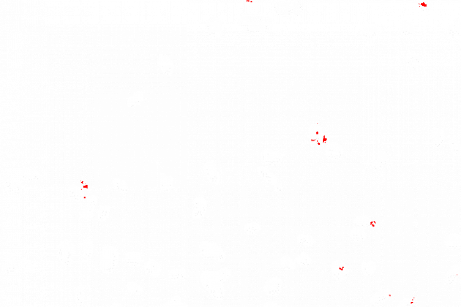
Tracking changes in cellular morphology and behavior in coordination with the surrounding cells. Cells with increasing area are marked in blue, cells with decreasing area in red. Length of individual cell borders, perimeter, area and percentile change compared to the previous image (Δ area, Δ perimeter) are tracked in real time.
Standard Deep Learning U-Net
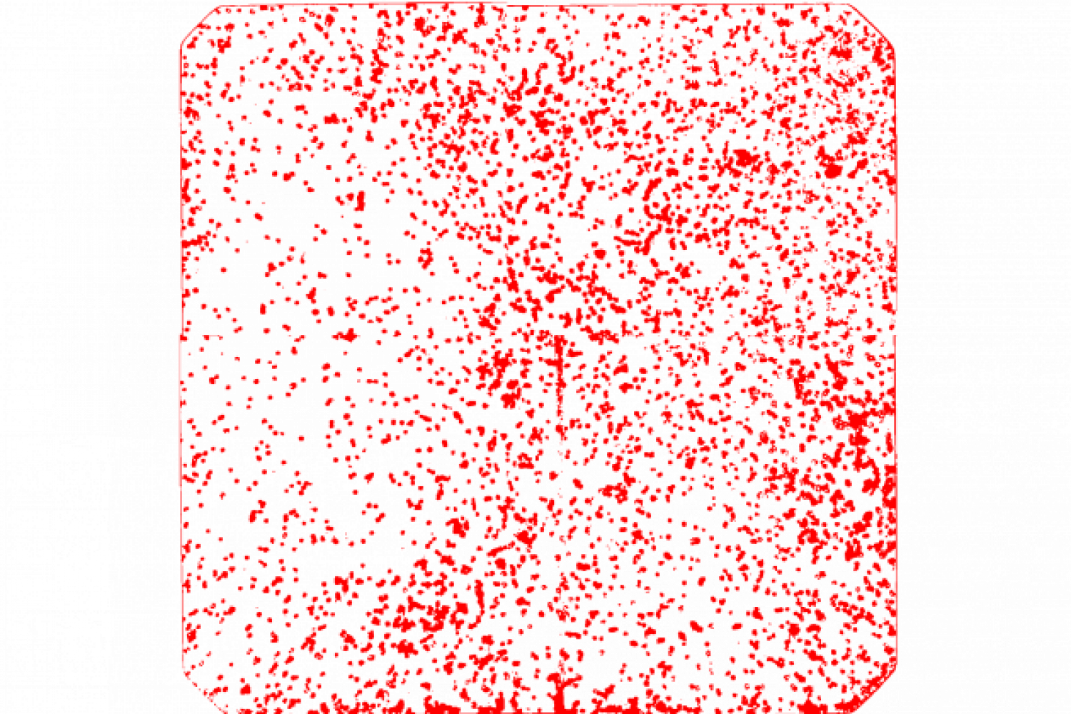
Simple intensity-dependent annotation of the cell boundaries compared to the original image.
<div class=”wpb_column vc_column_container vc_col-sm-6″>
<div id=”ut_inner_column_66cdac11202a4″ class=”vc_column-inner “>
<div class=”wpb_wrapper”>
<div class=”wpb_raw_code wpb_raw_html wpb_content_element”>
<div class=”wpb_wrapper”>
<u>AI networks developed in-house:</u>
<ul>
<li>+ Cell border gaps in the original image are closed intelligently</li>
<li>+ Automatically track the length of individual cell boundaries, changes in cell morphology and behavior in coordination with surrounding cells</li>
<li>+ Visual representation of selected morphological changes (area, circularity, behavior of neighboring cells, etc.)</li>
<li>+ Simple export of all statistical data to an Excel file</li>
</ul>
</div>
</div>
</div>
</div>
</div>
<div class=”wpb_column vc_column_container vc_col-sm-6″>
<div id=”ut_inner_column_66cdac1120656″ class=”vc_column-inner “>
<div class=”wpb_wrapper”>
<div class=”wpb_raw_code wpb_raw_html wpb_content_element”>
<div class=”wpb_wrapper”>
<u>Old U-Net based AI network:</u>
<ul>
<li>- Gaps in the original image are not closed, which affects the subsequent statistical analysis</li>
<li>- Tracking of cellular parameters difficult or only possible with additional programs</li>
<li>- No visual representation of selected changes in cell morphology</li>
</ul>
</div>
</div>
</div>
</div>
</div>
<h3>Counting and segmentation of cells in chamber slides</h3>


Detection and segmentation of nuclei and cytoplasm in cultured cells in chamber slides.
<h3>Segmentation of tumor and stromal cells</h3>


Segmentation of nuclei and cytoplasm in tumor cells and cytoplasmic regions in stromal cells. If nuclear staining is absent or non-specific, nuclei can be detected by nuclear “holes” in the cytoplasmic staining.
<h3>Analysis of the kidney</h3>


Segmentation of glomeruli and tubules using a deep learning model for Brightfield data.
<h3>Analysis of the kidney</h3>


Segmentation of glomeruli and their compartments using a tensor flow model for Brightfield data.
<h3>Cell counting</h3>


Segmentation and counting of cells.
<h3>Endoplasmic reticulum</h3>


The endoplasmic reticulum (ER) is a dynamic structure consisting of branched domains and tubular sections. Automatic segmentation opens up new possibilities for quantitative analysis and efficient visualization of this membrane structure at the ultra-structural level.
<h3>Scratch Assay</h3>


Analysis of samples with scratches and the change in cell area over time.
<h3>Internalization</h3>

Analysis of the insulin receptors inside and outside the cytoplasm and the changes over time.
<h3>Blood vessel analysis</h3>


Blood vessels can be labeled immunohistochemically. This enables blood vessel counting and analysis of blood vessel distributio
<h3>Colorectal carcinoma</h3>


Segmentation of Brightfield data with colorectal carcinoma using a deep learning model.
<h3>Proliferation</h3>
Segmentation of cells over time to measure cell proliferation. The analysis is performed on Label Free Data.

<h3>Real-time detection on microscopy devices</h3>
You look through the microscope and see markings of the objects relevant to you in real time. The detected objects are then automatically quantified.


<h3>Analysis of the lungs</h3>


Automated classification of tumors in non-small cell lung cancer (NSCLC) and Quantification of tumors/stromas.
<h3>Intracellular compartments in EM</h3>


Ultrastructural analysis of intracellular compartments using electron microscopy provides new insights into cellular fine structures and subcellular organelle interactions.